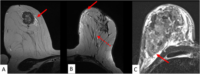
Fig. 5.
The pivotal role of T2-weighed images: oedema. Any asymmetric ipsilateral T2-weighted signal increase not due to the tumour itself, cysts or artefacts is referred to as “oedema” in A, B, and C. Different oedema patterns are distinguished such as perifocal (A), full red arrow), subcutaneous (B, full red arrow), prepectoral (C, full red arrow), and diffuse (B, dotted red arrow). Oedema is considered among the best evaluated predictive/prognostic criteria and was associated with high grade and nodal-positive cancers as well as disease recurrence (see also Table 1)

