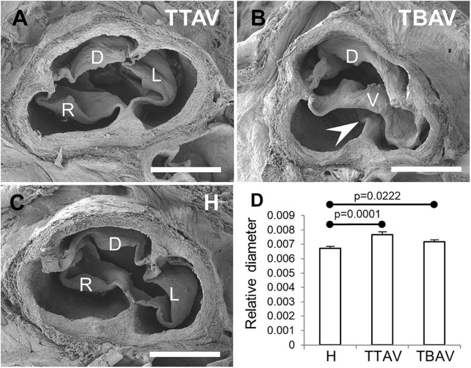
FIGURE 1.
Scanning electron micrographies of TAVs (A,C) and a BAV (B) of hamsters from the affected (A,B) and the control (C) strains. Cranial views. D, dorsal (non-coronary) leaflet; L, left leaflet; R, right leaflet; V, ventral leaflet. Arrowhead: raphe. Scale bar: 500 μm. (D) Analysis of the relative diameter of the ascending aorta of hamsters grouped according to the strain and the valve morphology. H, control strain; TBAV, bicuspid aortic valve from the affected strain; TTAV, tricuspid aortic valve from the affected strain. Only significant p-values are detailed.

