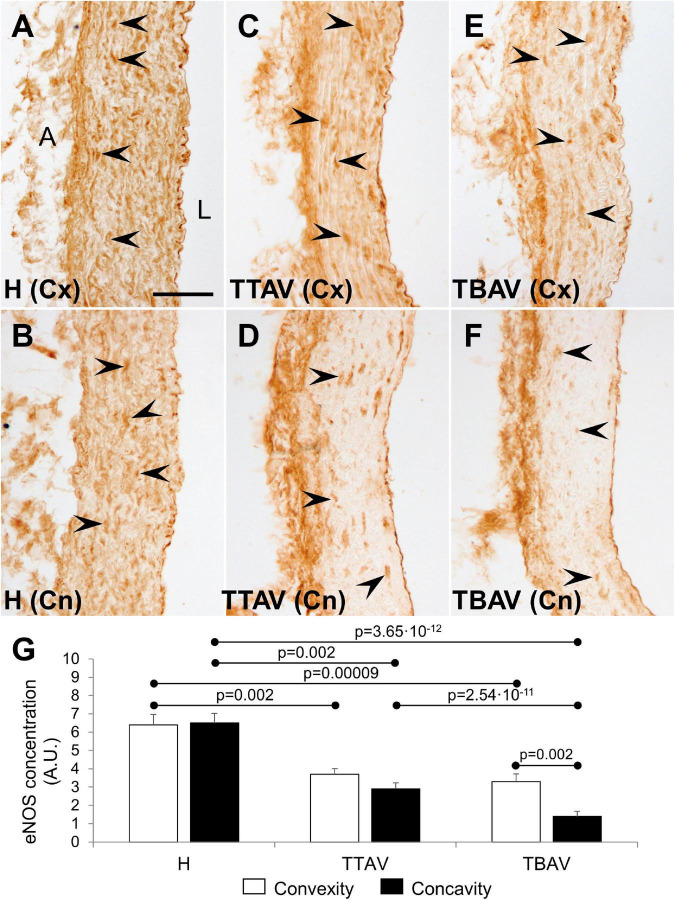
FIGURE 4.
Endothelial nitric oxide synthase immunoperoxidase in the convexity (Cx; A,C,E) and concavity (Cn; B,D,F) of the ascending aorta of hamsters from the control (A,B) and the affected (C–F) strains. All specimens showed strong immunoreactivity in the endothelium, close to the lumen (L), and the adventitia (A). Medial eNOS+ smooth muscle cells (arrowheads) were abundant and homogeneously distributed in animals from the control strain (A,B), but they were scarce and scattered in animals of the T strain (C–F), particularly at the aortic concavity (D,F). No signal was detected in the negative control (not shown). Scale bar: 50 μm. (G) Quantification of the aortic media area occupied by eNOS+ cells. Expression was reduced almost by halve in both the convexity and the concavity of animals of the T strain. Convexity and concavity showed similar eNOS expression except in animals with BAV, in which the reduction in the concavity was even more pronounced. H, control strain; TBAV, bicuspid aortic valve from the affected strain; TTAV, tricuspid aortic valve from the affected strain. Only significant p-values are detailed.

