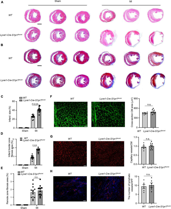
FIGURE 3.
The reduced expression of LEC-S1pr1 aggravates post-MI pathological cardiac remodeling. (A) Representative images of H&E staining of serial sections from hearts in WT and Lyve1-Cre-S1pr1flox/wt mice at 28 days after MI (n = 13). (B–E) Representative images of Masson’s Trichrome staining of serial sections from hearts in WT and Lyve1-Cre-S1pr1flox/wt mice at 28 days after MI (B), with quantification of the percentage of infarct size and cardiac fibrosis in left ventricle myocardium (C–E) (n = 13). (F) Representative images of WGA staining of hearts in WT and Lyve1-Cre-S1pr1flox/wt mice at 28 days after MI, with quantification of the cross-section cardiomyocyte area in hearts (n = 8). (G) Representative images of isolectin-B4 staining of hearts in WT and Lyve1-Cre-S1pr1flox/wt mice at 28 days after MI, with quantification of capillary density in post-MI myocardium (n = 8). (H) Representative images of Lyve-1 (LEC marker) staining of hearts in WT and Lyve1-Cre-S1pr1flox/wt mice at 28 days after MI, with quantification of capillary lymphatic density in post-MI myocardium (n = 6). Data are mean ± S.E.M. Scale Bars: (A,B), 2 mm; (F–H), 50 μm; n.s., no statistical significance.

