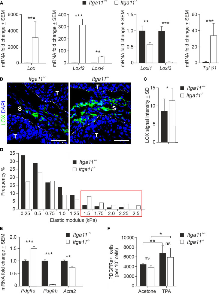
Figure 5.
Expression of LOX family members and CAF markers in Itga11−/− skin tumors. (A) RT-qPCR analysis of lysyl oxidase (Lox), LOX-like enzymes (Loxl1-4), and transforming growth factor beta-1 (Tgfβ1) in Itga11+/+ and Itga11-/- skin papillomas at week 20. The data are an average of ten samples (from different individuals) per genotype and normalized with endogenous Gapdh. (B) Representative immunofluorescence staining of LOX in Itga11+/+ and Itga11-/- papillomas. LOX signals are prominent in α11-deficient skin tumors and widely distributed within the tumor stroma. Scale bars, 50 μm. S, stroma; T, tumor. (C) Quantification of LOX immunofluorescence in Itga11+/+ and Itga11-/- papillomas. Twelve to 15 images from five tumors from five different individuals/genotype were quantified using Fiji ImageJ analysis software. (D) Tumor stiffness measurements by atomic force microscopy. Histograms of the Young’s elastic modulus (kPa) of the Itga11+/+ (n = 4) and Itga11-/- (n = 3) papillomas collected at weeks 20–25. (E) RT-qPCR analysis of fibroblast markers PDGFRα, PDGFRβ, and αSMA (Acta2) in Itga11+/+ and Itga11-/- papillomas collected at week 20. Data are an average of ten samples (from different individuals) per genotype. Values were normalized with Gapdh. (F) Numbers of Lin- PDGFRα+ cells in the acetone-treated normal and TPA-treated hyperplastic skin of the Itga11+/+ and Itga11-/- mice. In (A, C, E, F) *, p<0.05, **, p<0.01, ***, p<0.001. ns, not significant

