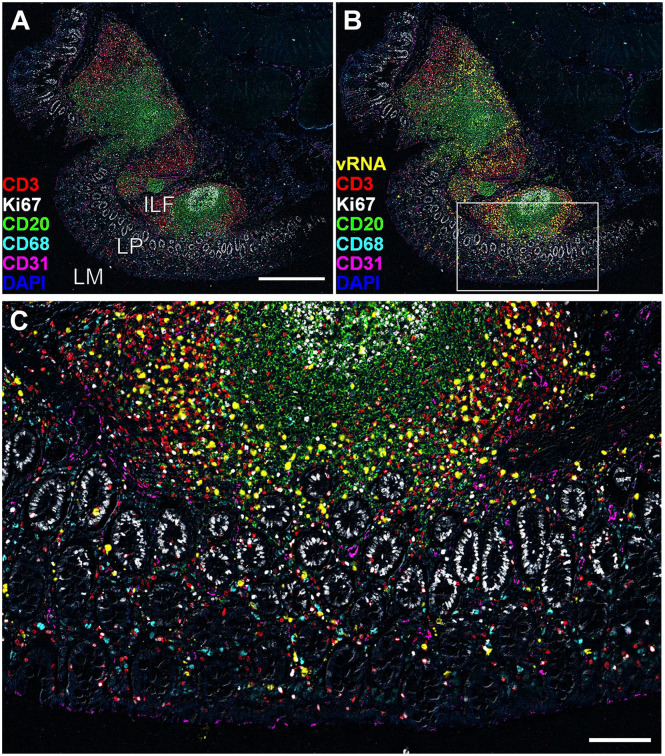
Figure 5.
Representative non-lymphatic tissue images detected using the Comb-CODEX-RNAscope where all pretreatments of CODEX and RNAscope were combined at the begin of the experiment. (A) The merged image of rectal tissue of a rhesus macaque infected with SIVmac251 (Rh4978, 10 dpi), showing CD3 (red), Ki67 (white), CD20 (green), CD68 (cyan), CD31 (magenta), and DAPI (blue). Scale bar equals 500 microns. (B) The image of A adds SIV vRNA (yellow). (C) The boxed area in image B was zoomed in, scale bar equals 100 microns. Abbreviations: CODEX, CO-detection by inDEXing; DAPI, 4′,6-diamidino-2-phenylindole; ILF, isolated lymphoid follicle; LP, lamina propria; LM, lumen; SIV, simian immunodeficiency virus.

