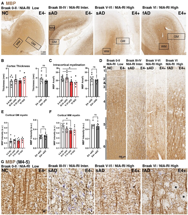
FIGURE 1.
Myelin basic protein-expressing fibers are reduced in sAD frontal cortex independent of APOE4 status. (A) Representative low-power micrograph of MBP immunohistochemistry on postmortem human frontal cortices from NC, sAD, and fAD. Braak stage of NFT or NIA-RI of NFT + NP are indicated on top left, APOE4 carrier status on top right; thicknesses of the GM and WM regions are indicated by rectangles. (B) No differences in the cortical GM thickness among groups by Braak stage or APOE4 status in sAD cohorts. (C) Myelinated (MBP+) areas of the GM are reduced in sAD and fAD with advanced Braak stages but are unaffected by APOE4 status in sAD. (D) Representative mid-power images of the cortical GM showing the 6 layers of myeloarchitecture (M1-6) (47). (E) No differences in the MBP intensity among groups by Braak stage or APOE4 status. (F) MBP intensity was reduced in sAD and fAD with Braak stage progression but unaffected by APOE4 status. (G) High magnification between myelin layer 4-5. Strong, aligned, and organized MBP+ myelin fibers were found in NC. The alignment and organization of MBP+ fibers arrangements were disrupted in sAD; aligned fibers were deflected by amyloid plaques or extracellular neurofibrillary tangles (asterisks) in fAD. Statistical analyses by one-way ANOVA among groups with Tukey post hoc test, or unpaired t-test for pairwise comparison (*p < 0.05).

