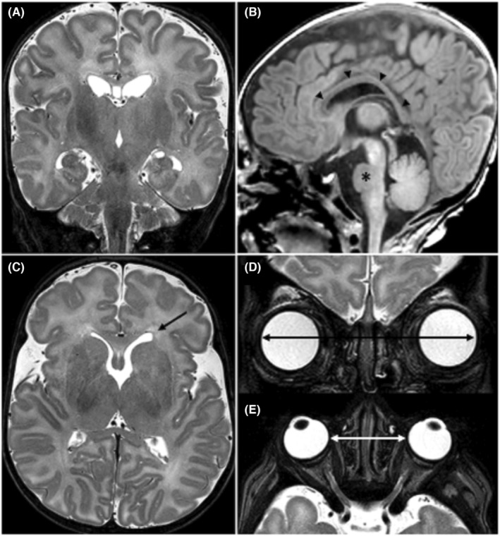
FIGURE 1.

MR imaging examination performed at nearly 2 months of age. Coronals (A, D) and axials (C, E) TSE T2‐weighted images and sagittal (B) 3D‐TFE T1‐weighted image. MR showed (A) a moderately increased brain volume, (B) a thin and dysmorphic corpus callosum (arrow heads) and a small pons (asterisk). (C) Mild asymmetry of the lateral ventricles with squared off left ventricular frontal horn (arrow). Increased (D) binocular and (E) interocular distances. Abbreviations: TSE, turbo spin echo; 3D‐TFE, three‐dimensional turbo field echo
