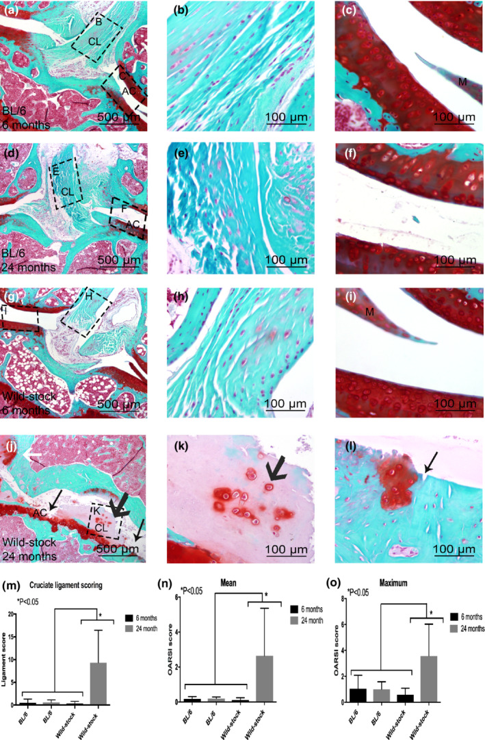
FIGURE 1.

Histological comparison between 6‐month‐old C57BL/6 (a–c), 6‐month‐old wild‐stock house mouse (d–f), 24‐month‐old C57BL/6 (g–i), and 24‐month‐old wild‐stock house (j–k) mouse with Safranin‐O knee joints. On histological images cruciate ligament (CL), articular cartilage (AC), and meniscus (M) is shown. An abnormal structure of cruciate ligaments with chondrocytic cell morphology and increased proteoglycan content around cells was observed (black wide arrow in j and k). Erosion to the calcified cartilage extending >75% of the articular surface (black arrow in j and l) and osteophyte formation and structural changes in meniscus were also observed (white arrows in j). Statistically significantly higher CL and OARSI scores were measured in 24‐month‐old wild‐stock house mice compared with 6‐, 24‐month‐old C57/BL6, and 6‐month‐old wild‐stock house mice (m–o). Data are means ± SEM.
