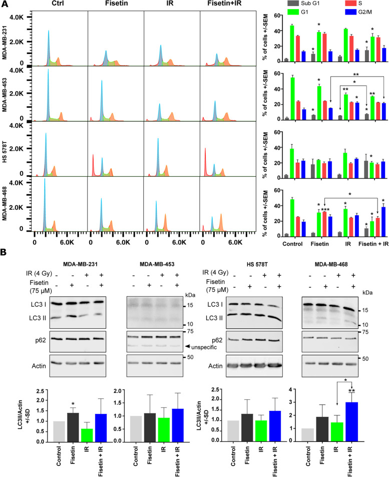
Fig. 6.
Fisetin in combination with irradiation does not affect cell cycle progression and autophagy. A log-phase cells were treated with fisetin (75 µM) for 24 h and irradiated with 4 Gy. Forty-eight hours after irradiation, cells were collected and fluorescence-activated cell sorting analysis was performed as described before [27]. The percentage of cells in different cell cycles as mean ± SD was calculated from at least 3 independent experiments and graphed. The asterisks indicate significant differences in individual treatment groups compared to the DMSO treated control (Ctrl) condition or between the arrows indicated conditions (*p < 0.05, **p < 0.01, ***p < 0.001; students t-test). B The cells were treated with a vehicle or fisetin (75 µM) for 65 h and mock irradiated or irradiated with 4 Gy. Protein samples were isolated 7 h after irradiation and subjected to SDS-PAGE. The level of LC3I/II and p62 were detected using Western blotting. The experiments were repeated at least 3 times and similar results were obtained. Actin was detected as the loading control. Bar graphs represent the mean densitometry of LC3-II to actin from at least 3 independent experiments normalized to 1 in control condition. The asterisks indicate significant differences in mean LC3-II/actin between the indicated conditions or compared to untreated control (*p < 0.05, **p < 0.01; students t-test)

