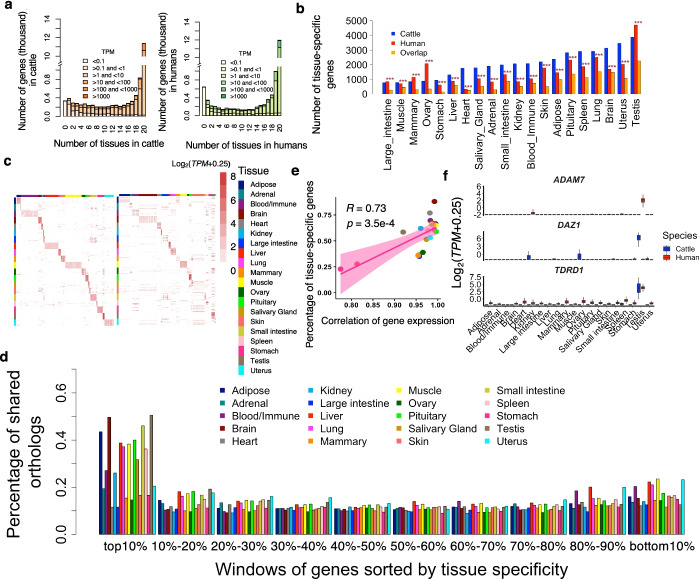
Fig. 2.
Comparison of tissue specificity of gene expression. a Gene expression levels and number of tissues in which genes were expressed (median TPM > 0.1) in cattle (left) and humans (right). b Number of tissue-specific genes (log2(fold-change) > 1.5 and FDR < 0.05) and their overlap across 20 tissues in humans and cattle. The overlap was tested using hypergeometric test. “***” represents FDR (Benjamini-Hochberg method corrected P-value) less than 1.0×10⁻³. c Expression profiles of top 10 tissue-specific genes that are detected in cattle among both cattle (left) and humans samples (right). Each row represents a gene and each column represents a sample from the corresponding tissue. The color represents log2-transformed expression value, i.e., log2(TPM+0.25). d Percentage of orthologous genes shared in each bin between humans and cattle. Genes were ranked (from largest to smallest) by degree (measured by −log10p) of tissue specificity, and then divided into ten bins (1731 genes per bin). e Spearman’s correlation between the percentage (%) of overlapping tissue-specific genes and gene expression correlation between humans and cattle across 20 tissues. Each dot represents a tissue. f Expression profiles of ADAM7 (human-specific testis gene), DAZ1 (cattle-specific testis gene), and TDRT1 (conserved testis gene)

