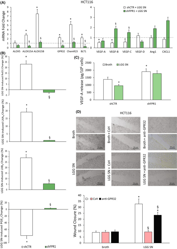
Fig. 6.

Dependence of Lactobacillus rhamnosus GG (LGG) supernatant (SN) effects on formyl peptide receptor 1 (FPR1) expression in colorectal carcinoma (CRC) cells. (A) ALOX5, ALOX15A, ALOX15B, GPR32, ChemR23, BLT1, VEGF‐A, VEGF‐B, VEGF‐C, VEGF‐D, Ang1 and CXCL1 mRNA fold change in HCT116 cells silenced for FPR1 (HCT116 shFPR1, three clones) or in control cells transfected with nontargeting short hairpin RNAs (shCTR cells, a mass population) upon treatment for 3 h with Lactobacillus rhamnosus GG (LGG) supernatant (SN)—1 : 30 titration. Data are represented as mean ± SD of five independent experiments. *P < 0.05 compared with the control (broth—dotted line) by Student's t test, § P < 0.05 compared with the relative control by Student's t test. (B) Proresolving and proinflammatory autacoid (RvD1, LXA4, LXB4, PGE2) release over control in HCT116 shCTR (a mass population) or shFPR1 (three clones) upon treatment for 12 h with LGG SN—1 : 30 titration. Baseline values of each mediator were in HCT116 shCTR cells: RvD1 128 ± 18 pg/106 cells, LXB4 41 ± 5 pg/106 cells, LXA4 492 ± 51 pg/106 cells and PGE2 98 ± 13 pg/106 cells. Baseline values of each mediator were in HCT116 shFPR1 cells: RvD1 68 ± 7 pg/106 cells, LXB4 18 ± 3 pg/106 cells, LXA4 346 ± 42 pg/106 cells and PGE2 214 ± 28 pg/106 cells. Data are represented as mean ± SD of changes over baseline levels obtained in five independent experiments. *P < 0.05 compared with the broth control by Student's t test, § P < 0.05 compared with the relative control by Student's t test. (C) VEGF‐A release in HCT116 shCTR (a mass population) or shFPR1 (three clones) cells treated with LGG SN—1 : 30 titration or the culture broth for 12 h. Data are represented as mean ± SD of five independent experiments. *P < 0.05 compared with shCTR broth by Student's t test. (D) Wound‐healing assay of HCT116 cells in the presence of LGG SN (1 : 30 titration) or the same dilution of culture broth for 12 h. Cells were pretreated or not for 30 min with CsH (800 nm) or a neutralizing anti‐GPR32 antibody (1 μg·mL−1). Representative photograms and a quantitative evaluation of the wound closure are shown. Scale bar 50 μm. Values represent the average of triplicate experiments ± SD. *P < 0.05 compared with broth alone by Student's t test. § P < 0.05 compared with the relative control by Student's t test. [Colour figure can be viewed at wileyonlinelibrary.com]
