
Fig. 7.
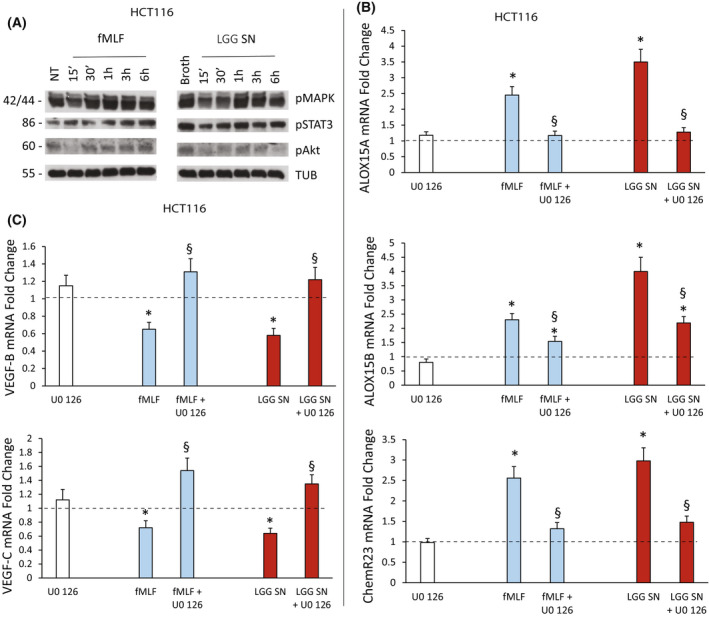
Signaling pathway activation upon fMLF or Lactobacillus rhamnosus GG (LGG) supernatant (SN) treatment of colorectal carcinoma (CRC) cells. (A) Activation of downstream signaling pathways in HCT116 cells induced by fMLF (10−9 m) or Lactobacillus rhamnosus GG (LGG) supernatant (SN) (1 : 30 titration) or the relative controls [not‐treated (NT) or broth alone, respectively]. Total cell lysates were prepared at various time points after stimulation in serum‐free medium. Immunoblots were probed with the indicated phosphospecific Abs. Antitubulin was used for normalization. The figure shows the results of a representative experiment from among three different preparations. (B) ALOX15A, ALOX15B and ChemR23 mRNA fold change in HCT116 cells treated with fMLF (10−9 m) or LGG SN (1 : 30 titration) for 3 h following or not cell preincubation with U0 126 (10 μm) for 30 min. Data are represented as mean ± SD of five independent experiments. *P < 0.05 compared with the negative control (dotted line) by Student's t test; § P < 0.05 compared with the relative control treatment by Student's t test. (C) VEGF‐B and VEGF‐C mRNA fold change in HCT116 cells treated with fMLF (10−9 m) or LGG SN (1 : 30 titration) for 3 h following or not cell preincubation with U0 126 (10 μm) for 30 min. Data are represented as mean ± SD of five independent experiments. *P < 0.05 compared with the negative control (dotted line) by Student's t test; § P < 0.05 compared with the relative control treatment by Student's t test. [Colour figure can be viewed at wileyonlinelibrary.com]