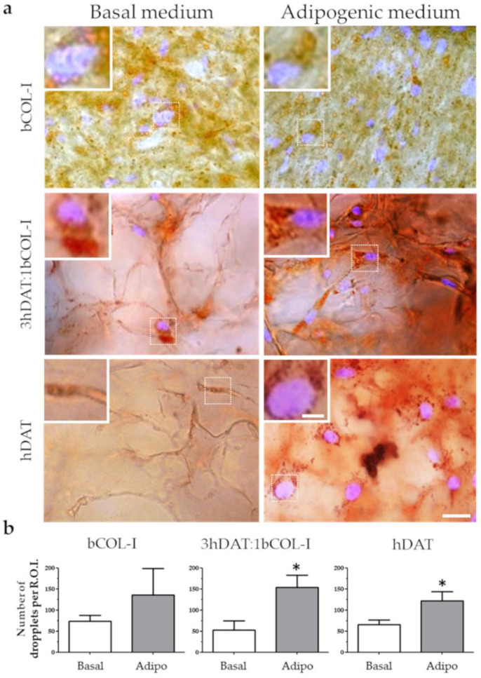
Figure 6.
Lipid deposition staining of hDPSCs cultured in solid foams with basal or adipogenic medium for 14 days in culture. (a) Images of Oil red (red) counterstained with DAPI. Upper row: bCOL-I, middle row: 3hDAT:1bCOL-I, and lower row: hDAT solid foams (scale bars represent 50 µm, inset 10 µm). (b) Quantification of the Oil Red stained droplets of the different culture conditions. Student’s t-test * p < 0.05.

