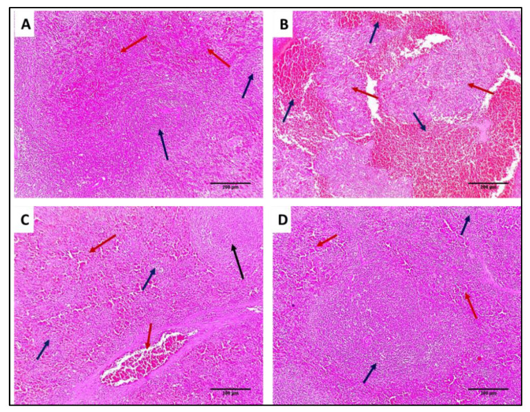
Figure 14.
H&E-stained sections: (A) Normal spleen showing normal-sized white pulp (lymphoid follicles) (blue arrows) with average-sized red pulp (blood sinusoids) (red arrows) (×100). (B) Spleen of group I showing marked congestion exhibiting dilated congested red pulp and areas of hemorrhage (blue arrows) with destructed white pulp (red arrows) (×100). (C) Spleen of group II showing mild congestion in the red pulp (blue arrows) with one preserved size of lymphoid follicle (black arrow) and other some atrophic lymphoid follicles (blue arrow) (×100). (D) Spleen of group III showing preserved white pulp (lymphoid follicles) (blue arrows) with normal red pulp (red arrows) (×100).

