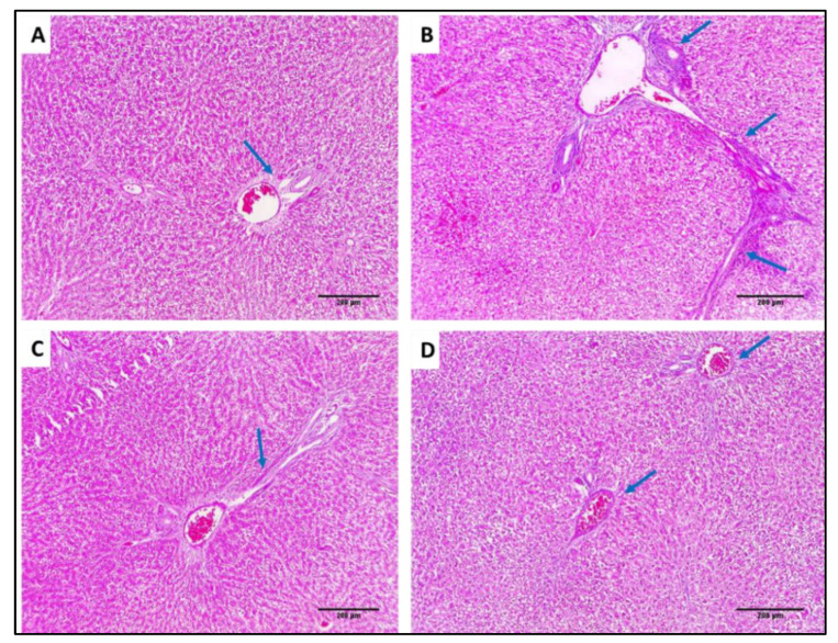
Figure 15.
Masson’s trichrome-stained sections: (A) Normal liver showing portal tract with a slight fibrous wall (blue arrow), and there is no fibrosis (×100). (B) Liver of group I showing the fibrous expansion of portal areas with a marked portal to portal bridging (blue arrows) (×100). (C) Liver of group II showing some fibrous expansion of portal areas (blue arrow) (×100). (D) Liver of group III showing few fibrous expansion of portal areas (blue arrow) (×100).

