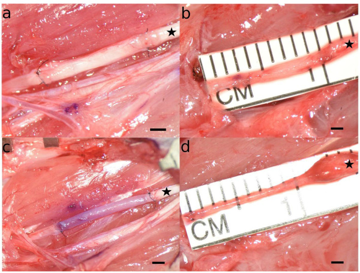
Figure 4.
Microscopic appearance of autologous nerve grafts (a,b) and muscle-in-vein conduits (c,d) immediately after nerve reconstruction (a,c) and at the timepoint of sacrifice twelve weeks after the initial surgery (b,d). Note that while the autologous neve grafts at WPO12 were comparable both in length and diameter to the originals grafts sutured to the stumps of the median nerve during the initial surgery, the muscle-in-vein-conduits appeared markedly thinner and stretched at the timepoint of sacrifice as compared to the initial surgery. Additionally, a prominent coaptation neuroma was observable at the proximal coaptation site in almost all the cases a muscle-in-vein conduit was used for nerve reconstruction. The proximal part of the reconstructed nerve is marked with an asterisk. Scale bar = 1 mm.

