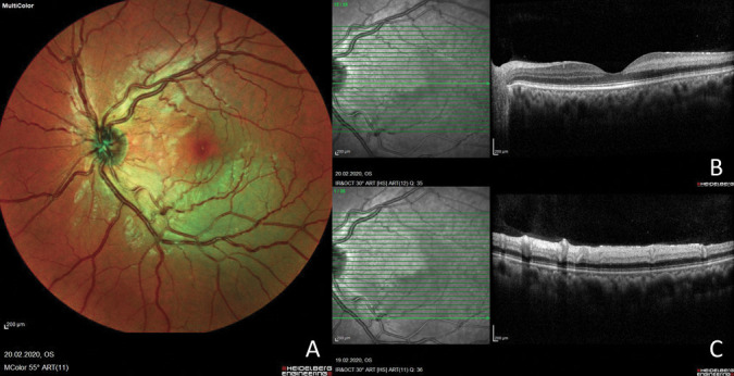
Fig. 1.

A. Multicolor fundus photograph of the left eye showing a deep retinal whitening area around the inferotemporal arcade. B. Optical coherence tomography (a horizontal slab on the fovea) of the left eye showing a hyperreflective band on the level of the INL consistent with PAMM. C. Spectral-domain OCT through the central area of the infarct displays ischemia and hyperreflectivity of both the middle and inner retinal layers.
