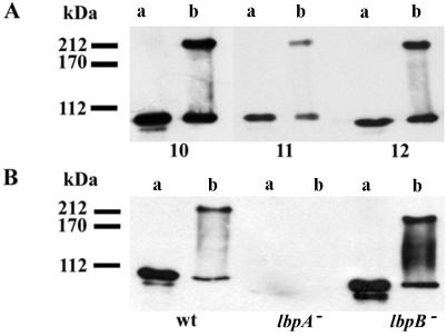
FIG. 2.
Western blot analysis of cell envelope proteins incubated at 100°C (lanes a) or 0°C (lanes b) before SDS-PAGE at 10 mA and 4°C. (A) Cell envelopes of strain BNCV probed with antisera (1:200 dilution) against synthetic LbpA peptides corresponding to loops 10, 11, and 12 in the topology model. (B) Cell envelopes of wild-type strain BNCV (wt), lbpA mutant CE1457 (8), and lbpB mutant CE1454 (12) probed with an LbpA-specific MAb. The binding of the primary antibody was monitored by incubation with a horseradish peroxidase-conjugated goat anti-mouse immunoglobulin antiserum (GAMPO; Jackson ImmunoResearch Laboratories, Inc.) and enhanced chemiluminescence (ECL) detection (Amersham). Molecular mass markers are indicated at the left.

