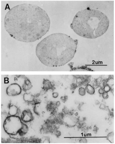
FIG. 1.
Electron micrograph of yeast spheroplasts (A) and vacuole-derived membranes (B). Spheroplasts and vesicles were fixed in 2% glutaraldehyde–0.5% osmium tetroxide, dehydrated with increasing concentrations of ethanol, and embedded in Epon-Araldite. Samples were stained with 1% uranyl acetate and Reynolds lead citrate and viewed and photographed with a Philips 300 electron microscope at 60 keV.

