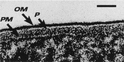
FIG. 3.
Freeze-substitution image of the E. coli K-12 cell envelope for direct comparison with Fig. 1. The periplasmic space is filled with periplasm (P) (the so-called periplasmic gel), and the peptidoglycan layer is not visible. The outer face of the outer membrane (OM) is densely stained, because the LPS retains its asymmetric location in this region of the bilayer and is more highly charged than the phospholipid on the inner face of the OM. The periplasm is bounded by the OM and the plasma membrane (PM). Bar = 25 nm. (Reprinted from reference 9.)

