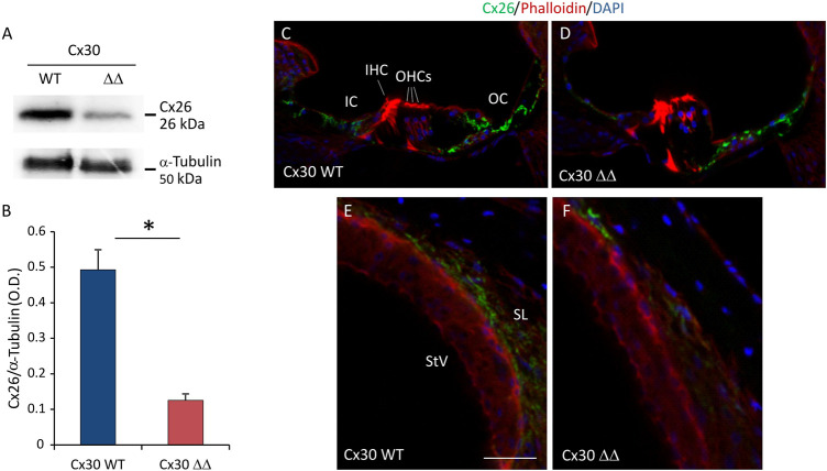
FIGURE 3.
Cx26 expression in Cx30 ΔΔ-aging cochleae. (A) Representative western blot immunoreactive bands showing the expression of Cx26 in cochlear lysates from Cx30 WT and Cx30 ΔΔ animals at 12 months of age. (B) Histograms (mean ± S.E.M.) represent optical density values normalized to α-tubulin. N = 8 cochleae/group. (C–F) Immunofluorescence analysis for Cx26 expression in Cx30 WT and Cx30 ΔΔ organ of Corti (C and D) and stria vascularis and spiral ligament (E and F) from 12 months of age cochleae. IHC: inner hair cell; OHCs: outer hair cells; OS: outer sulcus; IC: inner sulcus; StV: stria vascularis; and SL: spiral ligament. Scale bar: 50 μm. N = 4 cochleae/group. Experiments were performed in triplicate. Asterisks indicate significant differences between groups from Student’s t-test (*p < 0.05).

