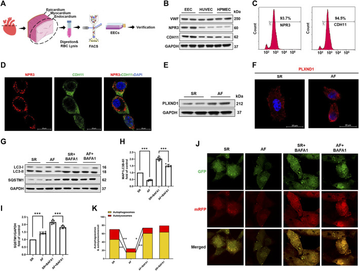
FIGURE 2.
Atrial fibrillation promotes PLXND1 expression and inhibits autophagy in endocardial endothelial cells. (A) Diagram of the EEC isolation from SR and AF mice. (B) Verification with specific markers (NPR3 and CDH11) in EECs compared to those in HUVECs and HPMECs by WB data. (C) Verification with specific markers (NPR3 and CDH11) in EECs compared to those in HUVECs and HPMECs by FACS. (D) Immunostaining data verifying the colocalization of EEC marker NPR3 (red) and CDH11 (green) in EECs. (E) Representative WB and quantification of PLXND1 in EECs isolated from SR and AF mice. (G–I) Representative WB and quantification of autophagy-related protein levels of EECs subjected to BAFA1 (100 nM) treatment or no treatment. (J) EECs from SR and AF mice were infected with a tandem GFP-mRFP-LC3 adenovirus for 24 h. Representative images show formation of puncta in different groups. Scale bar: 10 μm. (K) Quantitative analysis of yellow and free red puncta in merged images. The number of both yellow and free red puncta decreased in the EECs of AF mice compared to that in SR mice. (n = 10 cells per group; cells were isolated from three mice for one experiment and three independent experiments were performed; mean ± SD; *p < 0.05, **p < 0.01). EECs, endocardial endothelial cells; SR, sinus rhythm; AF, atrial fibrillation; HUVECs, human umbilical vein endothelial cells; HPMECs, human pulmonary microvascular endothelial cells; FACS, fluorescence-activated cell sorting; NPR3, natriuretic peptide receptor 3; CDH11, cadherin 11; WB, western blot.

