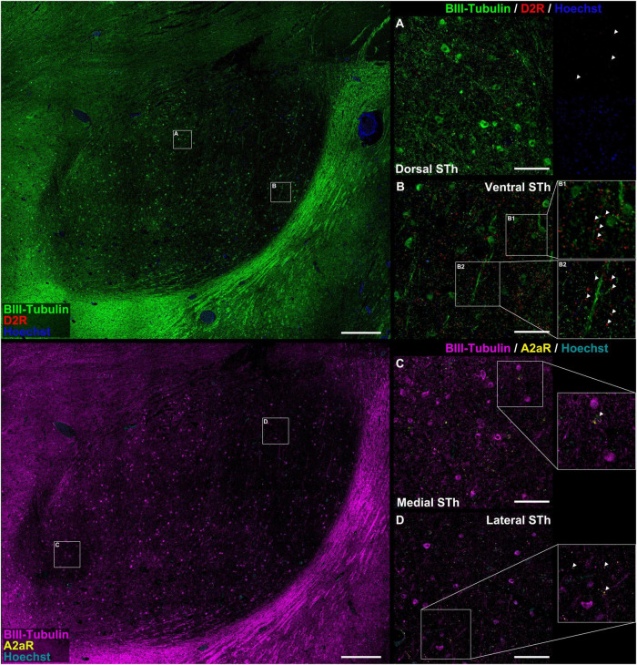
FIGURE 2.
Colocalization study between A2AR / D2R proteins and pan-neuronal marker β-III-tubulin. There appear to be significant differences in the distribution of D2 receptor immunofluorescent signal between the dorsal (A) and ventral (B) STh. In both instances, immunoreactivities mostly colocalized with β-III-tubulin neuritic structures at the level of dendritic spines [magnification in (B)]. In some occasions, D2R did not colocalize with β-III-tubulin, suggesting for the expression of the receptor also in non-neuronal cells. A2AR can be found as dot-like immunoreactivities colocalizing with β-III-tubulin positive neurites or as rare somatic cytoplasmic reactivities (C,D), even though non-β-III-tubulin colocalizing reactivities were also found.

