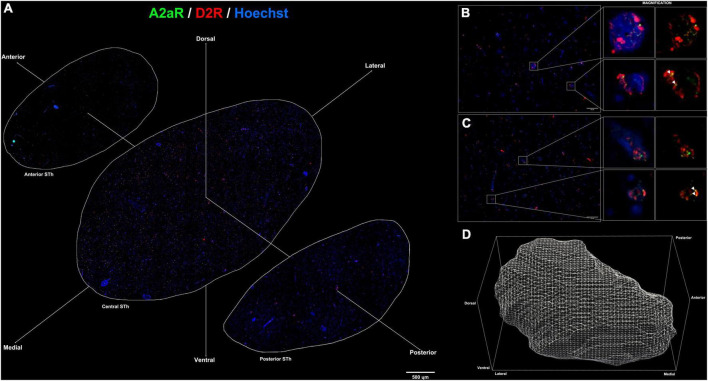
FIGURE 3.
(A) Immunofluorescent staining for A2AR (green) and D2R (red) throughout the rostro-caudal extent of the subthalamic nucleus reveals a peculiar topographical distribution. (B,C) colocalization of A2AR and D2R proteins within the subthalamic nucleus (white arrows). (D) 3D representation of the structure of the subthalamic nucleus following reconstruction of serial sections.

