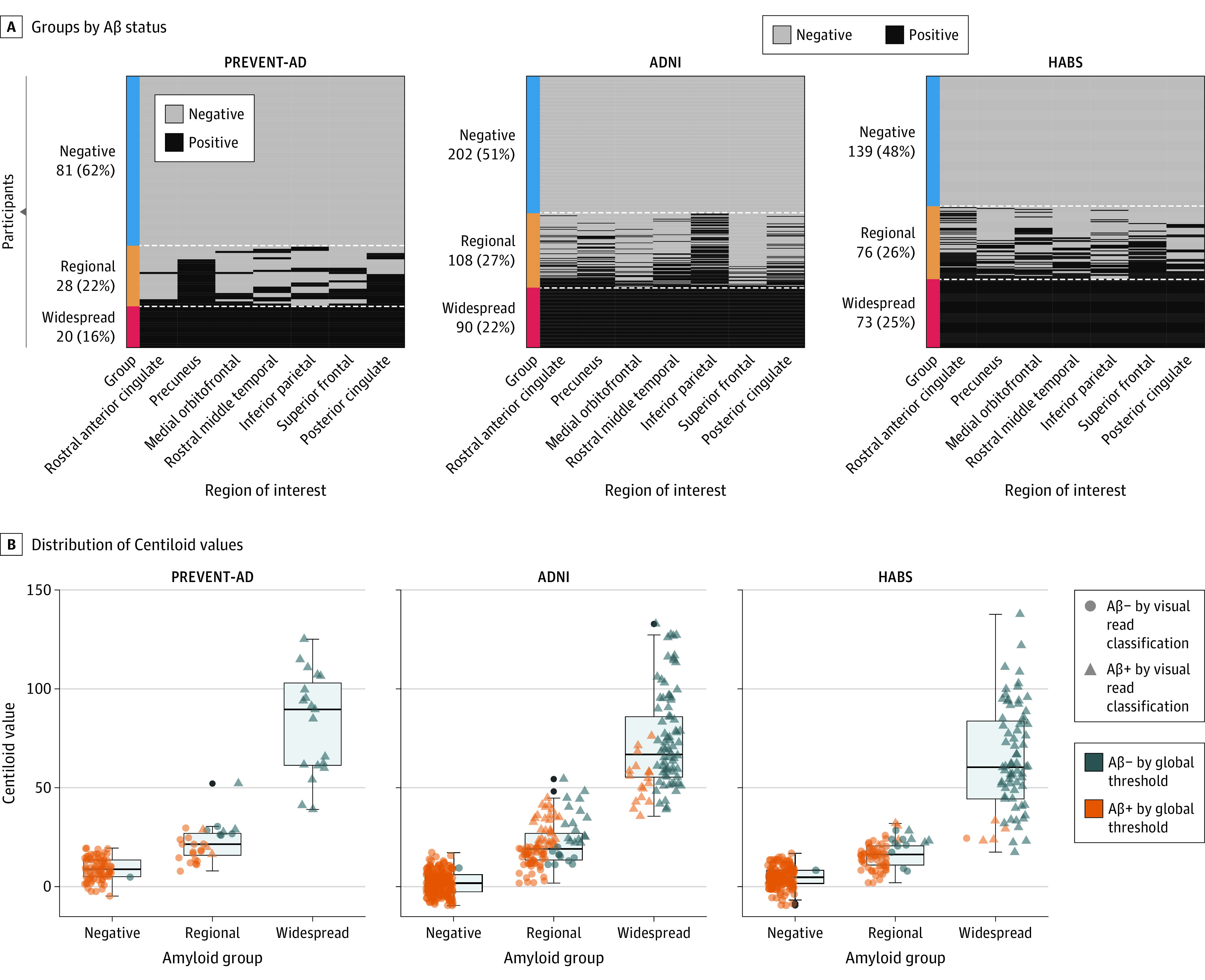
Figure 1. Defining the β-Amyloid (Aβ) Groups.

A, Individuals were separated into 3 groups based on their Aβ status in 7 cortical regions: rostral anterior cingulate, precuneus, medial orbitofrontal, rostral middle frontal, inferior parietal, superior frontal, and posterior cingulate. According to the region-specific positivity, individuals who were Aβ-positive in all 7 regions were classified as the widespread Aβ deposition group; those who were positive in 1 to 6 regions were included in the regional Aβ group; while those who fell below the cutoff in all the regions were considered as Aβ-negative. B, The boxplots represent the distribution of the Centiloid values of the 3 Aβ groups across the Aβ-negative, regional, and widespread groups in all cohorts. Different shapes for the data points indicate the visual read classification, and the color categorizes participants as Aβ+ or Aβ− based on quantitative binary amyloid index using previously established global thresholds for each cohort. ADNI indicates Alzheimer Disease Neuroimaging Initiative; HABS, Harvard Aging Brain Study; PREVENT-AD, Presymptomatic Evaluation of Experimental or Novel Treatments for Alzheimer Disease.
