Figure 3.

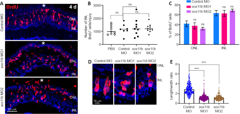
Sox11b knockdown does not affect MGPC cell number but causes changes in nuclear morphology at 4 dpi.
(A) BrdU immunofluorescence showed that MGPC formation was similar in control MO- and sox11b MO-treated retina at 4 dpi. White asterisks indicate sites of stab injury. (B) Quantification of BrdU+ cells at the injury site. (C) Proportions of BrdU+ cells located in the ONL or INL. (D) Higher-magnification images show differences in MGPC nuclear morphology, as indicated by white dotted lines, between control and Sox11b-depleted retinas at 4 dpi. (E) Quantification of the length/width ratio of MGPCs in the INL. Data are expressed as mean ± SEM (n = 3). Values were analyzed by one-way analysis of variance followed by Tukey’s post hoc test. ***P < 0.001. BrdU: 5-Bromo-2’-deoxyuridine; dpi: days post injury; GCL: ganglion cell layer; INL: inner nuclear layer; MGPC: Müller glia-derived progenitor cells; MO: morpholino; ns: not significant compared with control; ONL: outer nuclear layer.
