Figure 4.
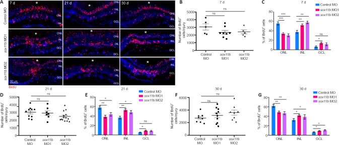
Sox11b loss-of-function alters MGPC distribution across retinal layers after 7 dpi.
(A) BrdU lineage tracing revealed greater proportions of Sox11b-depleted MGPCs in the inner retina at 7, 21, and 30 dpi. MGPCs were labeled with a pulse of BrdU at 4 dpi. White asterisks indicate sites of stab injury. (B, D, F) Quantification of the total number of BrdU+ cells at the injury site at indicated time points. (C, E, G) Quantification of the proportions of BrdU+ cells in the ONL, INL, or GCL at indicated time points. Data are expressed as mean ± SEM (n = 3). Values were analyzed by one-way analysis of variance followed by Tukey’s post hoc test. *P < 0.05, **P < 0.01, ***P < 0.001. BrdU: 5-Bromo-2’-deoxyuridine; DAPI: 4’,6-diamidino-2-phenylindole dihydrochloride; dpi: days post injury; GCL: ganglion cell layer; INL: inner nuclear layer; MO: morpholino; ns: not significant compared with control MO; ONL: outer nuclear layer.

