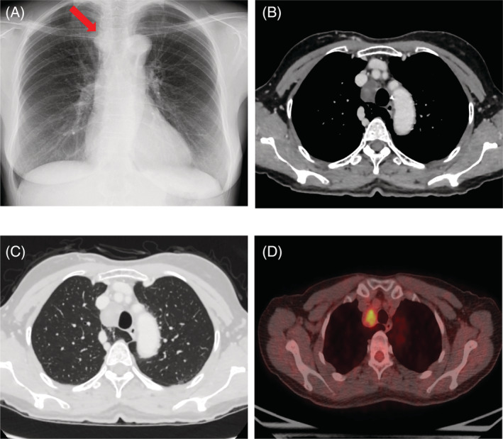
FIGURE 1.

(A) Chest X‐ray revealed a mass in the upper mediastinum. (B) contrast‐enhanced CT revealed a 3‐cm solid lesion with a smooth surface and poor contrast effect slightly excluding the right wall of the trachea, but there was no tendency to infiltrate the surrounding tissue. (C) This case had an azygos lobe near the tumour. (D) 18F‐fluorodeoxyglucose positron emission tomography (FDG‐PET) showed a slight uptake in the tumour
