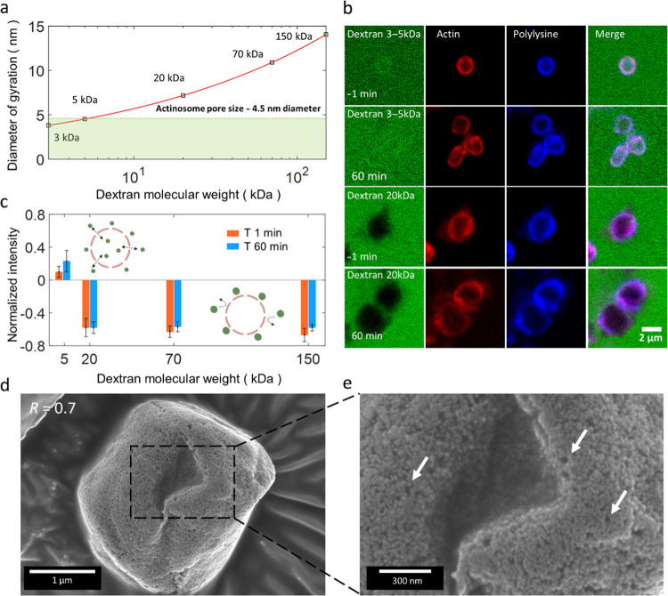
Figure 3.
Actinosomes are porous and permeable to small molecules. (a) Diameter of gyration (Dg) of dextran molecules as a function of their molecular weights (M). The red line follows the equation Dg = 2.64 × M0.33. (b) Confocal images showing the permeability of actinosomes (R = 0.7) to dextran molecules of different sizes, immediately (t0) and 1 h (t60) after incubation. Low-molecular-weight dextran (3–5 kDa) readily diffuses inside actinosomes, whereas high-molecular-weight dextran (20 kDa) is excluded from the actinosome. (c) Graph showing the normalized intensity (Iinside – Ioutside)/Ioutside of FITC-dextran at t0 (red) and t60 (blue). Positive values indicate dextran diffusion into the actinosomes, while negative values indicate impermeability to dextran. Error bars indicate standard deviations. (d) Scanning electron microscopy images of actinosomes (R = 0.7) appear as slightly crumpled spheres, similar to fluorescence images. (e) A zoom-in reveals a rough, unstructured, porous surface. Several sub-μm-sized pores are clearly visible and indicated with arrows. Error bars indicate standard deviations.

