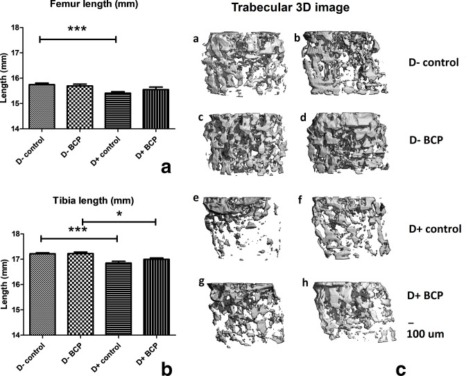
Fig. 2.
Relative length of tibia and femur bones and 3D image reconstruction of trabecular bone in the femur. Mice were fed a vitamin D (VD)-sufficient or VD-deficient diet and received either control or ß-caryophyllene (BCP) treatment, as described in the legend to Figure 1. a) Femur length (mm). Control groups: *p < 0.001. b) Tibia length (mm). Control groups: *p < 0.001; BCP groups: *p < 0.050. a) and b) show a statistically significant shortening of tibia and femur in mice fed a vitamin D-deficient diet. c) 3D image reconstruction of trabecular bone from 100 micro-CT slices each of femur bones from the various groups of mice. Two samples are shown in each treatment group.

