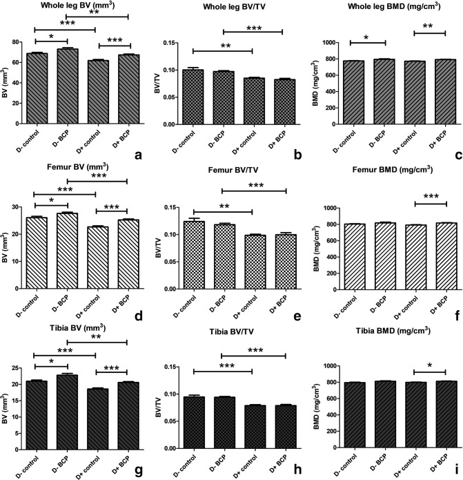
Fig. 3.
Micro-CT (μCT) of whole leg, femur, and tibia reveals the properties of bone in the presence or absence of vitamin D in mice treated with ß-caryophyllene (BCP). Mice were fed a vitamin D-sufficient or -deficient diet and received control or BCP treatment, as described in the legend to Figure 1. Bone samples from the whole leg, femur, and tibia of mice from each treatment group were subjected to μCT using a μCT 40 desktop μCT scanner (Scanco Medical, USA). a), b), and c) reflect the results of scans of whole leg; d), e), and f) show scans of femur; and g), h), and i) show scans of tibia. Bone volume (BV) is depicted in panels a), d), and g); bone volume/total volume (BV/TV) is shown in panels b), e), and h); bone mineral density (BMD) is depicted in panels c), f), and i). *p < 0.050; **p < 0.01; ***p < 0.001.

