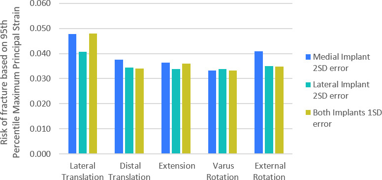
Fig. 3.

Bar graph showing the percentage difference in risk of fracture as the positions of the isolated medial unicondylar knee arthroplasty (UKA-M) and isolated lateral unicondylar knee arthroplasty (UKA-L) components were varied from the planned bi-unicondylar knee arthroplasty based on the typical accuracy achieved with manual instruments. A 2 standard deviation (SD) variation of the UKA-M implant only (blue), 2 SD variation in the UKA-L only (green), and 1 SD variation of both implants (yellow) are shown. In all cases the variation acts to narrow the bone island and/or make it taller.
