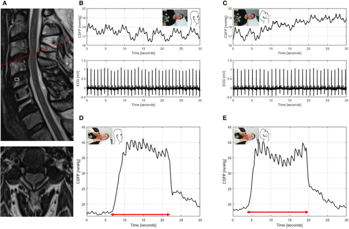
Figure 6.
T2-weighted MRI of the cervical spine for ID7 (patient with spinal canal stenosis of unknown significance, previously underwent surgery for degenerative cervical myelopathy, mJOA:9) (A). Sagittal and axial images showing spinal canal narrowing with spinal cord induration at level C3/4. CSFP and electrocardiogram (ECG) are shown during resting state in neutral head position with baseline CSFP of 17.4 mmHg and cardiac-driven CSFP peak-to-trough amplitude (CSFPp) of 0.5 mmHg (B), and in head reclination with baseline CSFP of 18.5 mmHg and CSFPp of 0.4 mmHg (C). CSFP is plotted during the Queckenstedt's test in neutral head position (rise, 17.7 mmHg) (D), and in head reclination (rise, 18.7 mmHg) (E).

