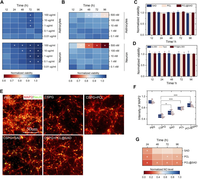
FIGURE 2.
In vitro cell viability and neuroprotective effect of PCL@SAD. (A,B) The effect of SAD nanoparticles (A) and PCL (B) on the neuronal and astrocyte viability (n = 6). (C,D) SAD nanoparticles, PCL, and PCL@SAD did not affect the viability of astrocyte (C) and neurons (D) (n = 6). The concentration of PCL was fixed at 10 nm; the amount of bare SAD nanoparticles was equal to that of PCL@SAD. (E) Representative images of axonal regeneration with different formulations after the stimulation with CSPG. The concentration of PCL was fixed at 10 nm; the amount of bare SAD nanoparticles was equal to that of PCL@SAD. (F) PCL@SAD, PCL, and SAD all improved the intensity of MAP2, when compared with the PBS group (n = 6). (G) The effect of PCL@SAD on the release of NO from neurons stimulated by lipopolysaccharide (n = 6). The concentration of PCL was fixed at 10 nm; the amount of bare SAD nanoparticles was equal to that of PCL@SAD. * p < 0.05, ** p < 0.01, and *** p < 0.001.

