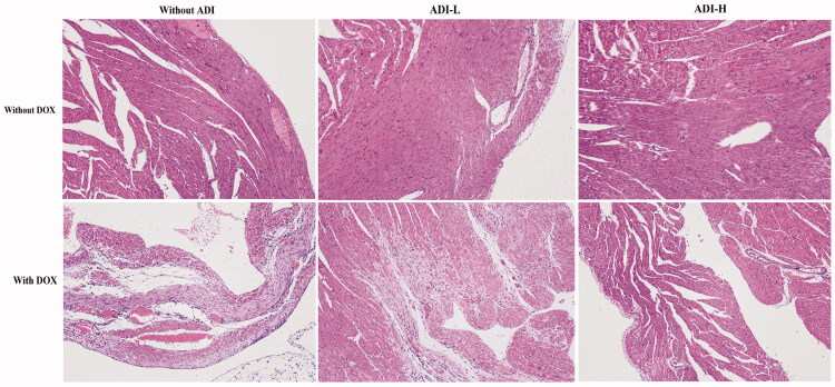
Figure 2.
H&E staining images of heart tissues in mice treated with DOX (0.03 mg/10 g) and/or ADI (0.1 and 0.2 mL/10 g) (100× magnification). Cells in the control group were in good condition and neatly arranged. In the DOX treatment group, myocardial cell disorders and increased cellular gaps were observed. In the ADI treatment group, no significant changes in cell morphology were seen. In the DOX combined with ADI group, injuries to the cells were slightly reduced.

