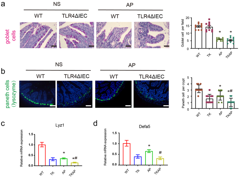
Figure 3.

Changes in intestinal cells in TLR4ΔIEC mice during acute pancreatitis.
(a) Representative images of intestinal goblet cells stained with PAS (400× magnification). (b) Representative images of intestinal Paneth cells stained with lysozyme by immunofluorescence (200× magnification). (c) Intestinal mRNA expression of Paneth-related genes (Lysozyme1 and Defensin-alpha 5). Data are provided as the mean ± SEM (n = 6 per group). *means p< .05 vs WT, #means p< .05 vs AP.
