
Figure 4.
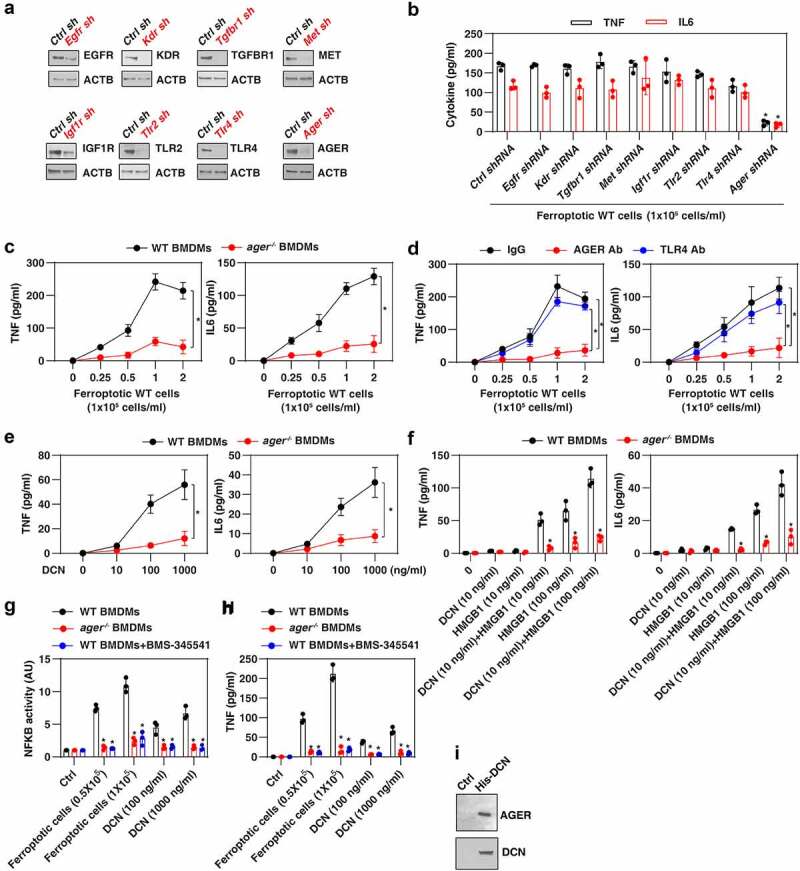
AGER mediates the response to DCN. (A) Western blot analysis of protein expression by BMDMs after transfection with the indicated shRNAs. (B) ELISA analysis of TNF and IL6 release in the indicated gene knockdown BMDMs following treatment with ferroptotic MEFs (n = 3 biologically independent samples; *P < 0.05 versus control shRNA group, one-tailed t test; data are presented as means ± SD). (C) Lack of Ager in BMDMs blocks ferroptotic cell-induced the production of the pro-inflammatory TNF and IL6 cytokines (n = 3 biologically independent samples; two-way ANOVA with Tukey’s multiple comparisons test; data are presented as means ± SD). (D) Anti-AGER neutralizing antibody (Ab; 1 mg/ml), but not anti-TLR4 neutralizing antibody (1 mg/ml), inhibits ferroptotic cell-induced TNF and IL6 release in BMDMs (n = 3 biologically independent samples; *P < 0.05, two-way ANOVA with Tukey’s multiple comparisons test; data are presented as means ± SD). (E, F) Lack of Ager in BMDMs inhibits DCN-induced TNF and IL6 release in the absence or presence of HMGB1 (n = 3 biologically independent samples; *P < 0.05 versus WT group, two-way ANOVA with Tukey’s multiple comparisons test; data are presented as means ± SD). (G, H) Analysis of NFKB activity and TNF release in the indicated BMDMs following treatment with ferroptotic cells or DCN in the absence or presence of the NFKB pathway inhibitor BMS-345541 (n = 3 biologically independent samples; *P < 0.05 versus WT group, two-way ANOVA with Tukey’s multiple comparisons test; data are presented as means ± SD). (I) His-tag affinity pull-down analysis of the binding of DCN to AGER.