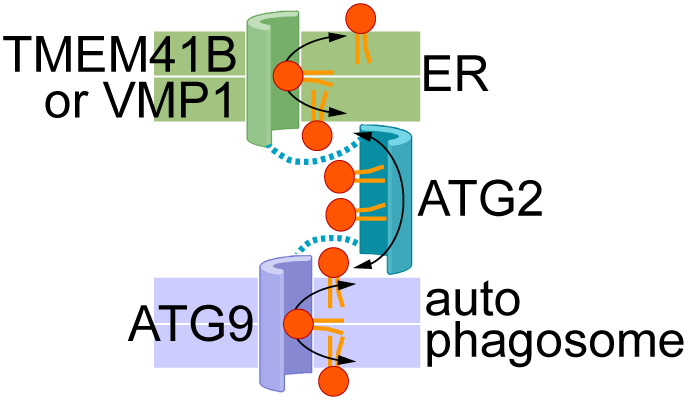
Figure 1. Proposed cooperation between scramblases in the ER (TMEM41B, VMP1) and nascent autophagosomal membrane (ATG9), and the bridging lipid transport protein ATG2.

The membrane bilayers are shown as coloured slabs (a white line separates the two halves of each bilayer). Phospholipids are shown generically with a red headgroup and orange acyl chains. Docking of ATG2 to the scramblases is indicated by the dotted lines. The transport proteins are shown according to the credit card model27, with a polar groove in the case of scramblases to accommodate lipid headgroups, and a hydrophobic groove in ATG2 to accommodate lipid tails. Bidirectional flow of lipids is shown by double-headed arrows so that lipids in both leaflets of both membranes are equilibrated by the scramblases and inter-bilayer exchange across the cytoplasm. Lipid transport may be effectively one-directional, with lipids being synthesized in the ER (‘source’) and consumed through expansion of the autophagosomal membrane (‘sink’) (see ‘Open Questions’ section ‘What drives lipids to move towards the acceptor membrane, i.e., the newly forming autophagosome membrane’ for details).
