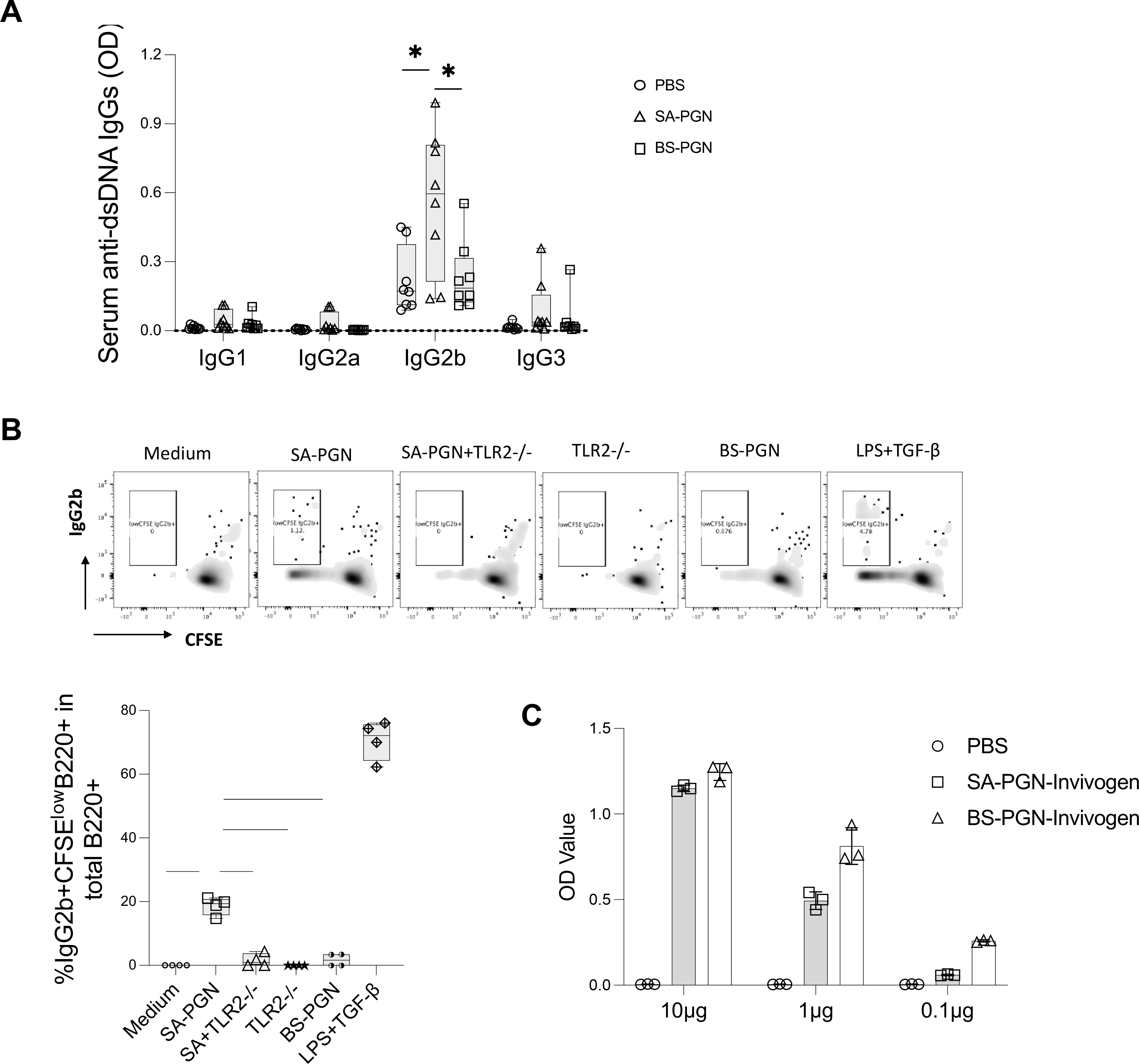
Figure 4.

IgG2b CSR mediated by S. aureus PGN. (A) C57/B6 mice were treated with S. aureus or B. subtilis PGNs via i.p. injection twice per week for 8 weeks. At the end of study, subclasses of serum anti-dsDNA IgG were evaluated in mice at 14 weeks of age. (B) B cells were isolated from spleen of untreated C57/B6 mice, cultured with basal medium, LPS plus TGF-β1 (a positive control), S. aureus (SA) or B. subtilis (BS) PGN in the presence of absence of TLR2 mAb or isotype Ab for 72 h. The percentages of proliferating IgG2b+ B cells (%CFSElowIgG2b+ in IgG2b+B220+ cells) were calculated. (C) The TLR2 induced activity by S. aureus and B. subtilis PGN (P = 0.25). Statistical analysis used non-parametric Mann-Whitney U tests.
