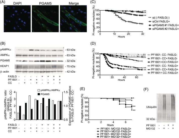
Figure 7.

Effect of PF1801 on AMPK‐PGAM5 pathway in FASLG‐mediated myotube necroptosis. (A) Immunofluorescence staining against PGAM5 (green) in myotubes. Nuclei were counterstained with DAPI (blue). Scale bar indicates 10 μm. (B) The protein expression of pAMPKα, AMPKα, PGAM5, KEAP1, and β‐actin evaluated with western blotting in the myotubes pretreated with PF1801 and/or CC and then treated with FASLG for 12 hours. The relative protein expression in western blot was analysed with ImageJ software. (C) The viability of myotubes transfected with scrambled siRNA (siCtrl: n = 97) or siRNA specific for Pgam5 (siPGAM5#1: n = 197, siPGAM5#2: n = 215) and treated with FASLG and that of myotubes without transfection nor FASLG treatment (siRNA‐FASLG−: n = 123). Log‐rank test, followed by Holm–Sidak multiple comparisons. **P < 0.01. (D) The viability of myotubes treated with PF1801, CC, and/or FASLG (n = 144; PF1801− CC− FASLG−, 152; PF1801− CC+ FASLG−, 154; PF1801+ CC+ FASLG+, 116; PF1801− CC− FASLG+, 169; PF1801− CC+ FASLG+, 157; PF1801+ CC− FASLG+). Log‐rank test, followed by Holm–Sidak multiple comparisons. **P < 0.01. (E) The viability of myotubes pretreated with PF1801 and/or MG132 and then treated with FASLG (n = 136; PF1801− MG132− FASLG+, 131; PF1801+ MG132− FASLG+, 151; PF1801− MG132+ FASLG+, 173; PF1801+ MG132+ FASLG+). Log‐rank test, followed by followed by Holm–Sidak multiple comparisons. *P < 0.05, **P < 0.01. (F) Immunoblotting analysis of ubiquitin in immunoprecipitated PGAM5 from myotubes treated with PF1801 and/or MG132. (B–F) Data represent three independent experiments.
