Figure 4.
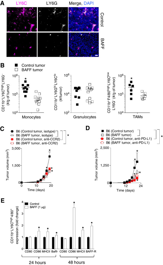
PD-L1 and monocytes are functionally important for the BAFF-mediated reduction in tumor growth. A–D, C57BL/6 mice were inoculated subcutaneously with 5 × 10⁵ of BAFF-expressing or control cells and tumors were analyzed as indicated at day 13. A, LY6C and LY6G expression in tumors was assessed using fluorescent IHC (representative images of n = 5–7 mice are shown). Scale bar, 50 μm. B, Numbers of monocyte, granulocyte, and TAMs infiltrates in tumors were analyzed using FACS (n = 6–10). C, C57BL/6 (B6) mice were treated with monocyte depleting antibody (anti-CCR2) and tumor growth was followed (n = 7–8, pooled from two independent in vivo experiments). D, C57BL/6 (B6) mice were treated with anti-PD-L1 antibody and tumor growth was followed (n = 4–5). E, Bone marrow–derived inflammatory monocytes were treated with 1 μg of BAFF protein for 24 hours 4 days post-isolation from the bone marrow. Expression of MHCII, CD86, CD80, and BAFF-R was analyzed using FACS (n = 4). Error bars in all experiments indicate SEM. *, P < 0.05 as determined by a Student t test (unpaired, two-tailed) or a two-way ANOVA with a post hoc test.

