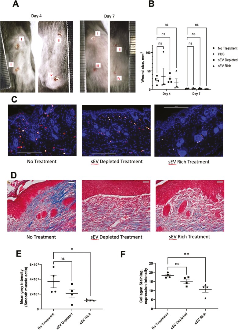
Figure 5.
Four identical wounds were created on the back of each animal and treatments were added for 4 days (A; I—no treatment, II—PBS, III—sEV depleted treatment, IV—sEV containing treatment). OMLP-PCL sEV treatment on mouse wounds demonstrates no statistically significant effect on wound size at 4 days (B). However, OMLP-PCL sEV treatment results in a reduction in α-SMA immunofluorescence and collagen deposition in vivo. Merged images showing nuclear DAPI (blue) staining and α-SMA (red) staining for non-treated, OMLP-PCL sEV depleted treatment, OMLP-PCL sEV treatment (C) and quantification of fluorescent α-SMA (E). Scale bar = 200 μm n = 4 wounds per group. Masson’s trichrome staining reveals collagen deposition in wounds from non-treated, OMLP-PCL sEV depleted treatment, OMLP-PCL sEV treatment (D) and quantification of collagen staining (F). n = 4 wounds per group, *P < .05, **P < .01, Scale bar = 30 μm.

