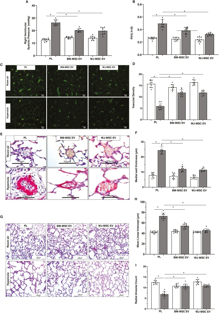
Figure 3.
BM and WJ-MSC EVs have similar effects in BPD and PH. Both IT BM-MSC and WJ-MSC EVs significantly reduced (A) right ventricular systolic pressure and (B) weight ratio of the right ventricle to left ventricle + septum (RV/LV+S) but the reduction in RV/LV+S was greater in 2 week old hyperoxia exposed rats who received WJ-MSC EVs. (C) Representative lung sections stained with Von Willebrand Factor showing increased vessels in both IT hyperoxia BM-MSC and WJ-MSC EV treated groups. Original magnification 10×. (D) Similar improvement in lung vascular density in both IT hyperoxia BM-MSC and WJ-MSC EV groups. (E) Lung sections stained with α-smooth muscle actin demonstrating decreased pulmonary vascular remodeling in hyperoxia-exposed rats treated with IT BM-MSC or WJ-MSC EV. Original magnification 40×. (F) Reduced medial wall thickness in hyperoxia-exposed rats treated with IT BM-MSC or WJ-MSC EVs. (G) H&E-stained lung sections demonstrating improved alveolar structure in hyperoxia-exposed rats that received IT BM-MSC or WJ-MSC EV. Original magnification 10×. Inset is 40×. Morphometric analysis showing (H) decreased mean linear intercept and (I) increased radial alveolar count in IT BM-MSC or WJ-MSC EV treated hyperoxia rats. Data are presented as mean ± SEM; N = 9/group. *P < .05; room air placebo (PL) vs. hyperoxia PL or hyperoxia PL vs. hyperoxia BM-MSC or WJ-MSC EV. #P < .05, hyperoxia BM-MSC EV vs. hyperoxia WJ-MSC EV. Room air: open bar; hyperoxia: gray bar.

