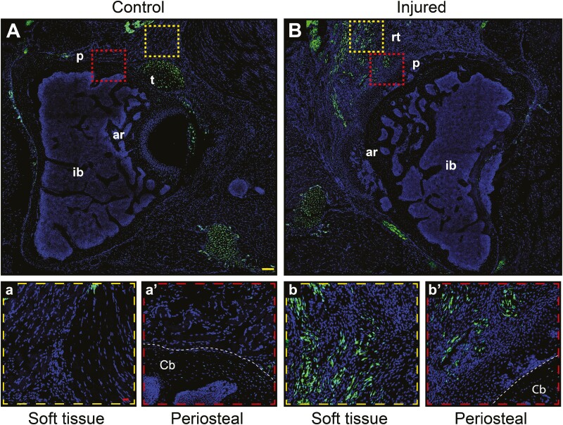
Figure 7.
Scleraxis expressing cells mark the areas that give rise to the heterotopic bone. Scleraxis (Scx)-GFP reporter animals underwent induction of heterotopic ossification (HO) by acetabular reaming, followed by analysis at 3 weeks post-operatively. Scx-GFP+ cells appear green. (A) Axial section taken from an uninjured hip (A-a’) with high magnification pictures showing more in detail the soft tissue (a) and periosteal (a’) areas. (B-b’) Axial section obtained 3 weeks after HO induction, demonstrating heterotopic bone in the soft tissue and periosteal periacetabular areas (b-b’). Abbreviations: p, periosteum area; t, tendon; ar, acetabular roof; ib, iliac bone; rt, reactive tissue; cb, cortical bone. DAPI nuclear counterstain appears blue. Yellow scale bar: 100 μm. Red scale bars: 20 μm.

