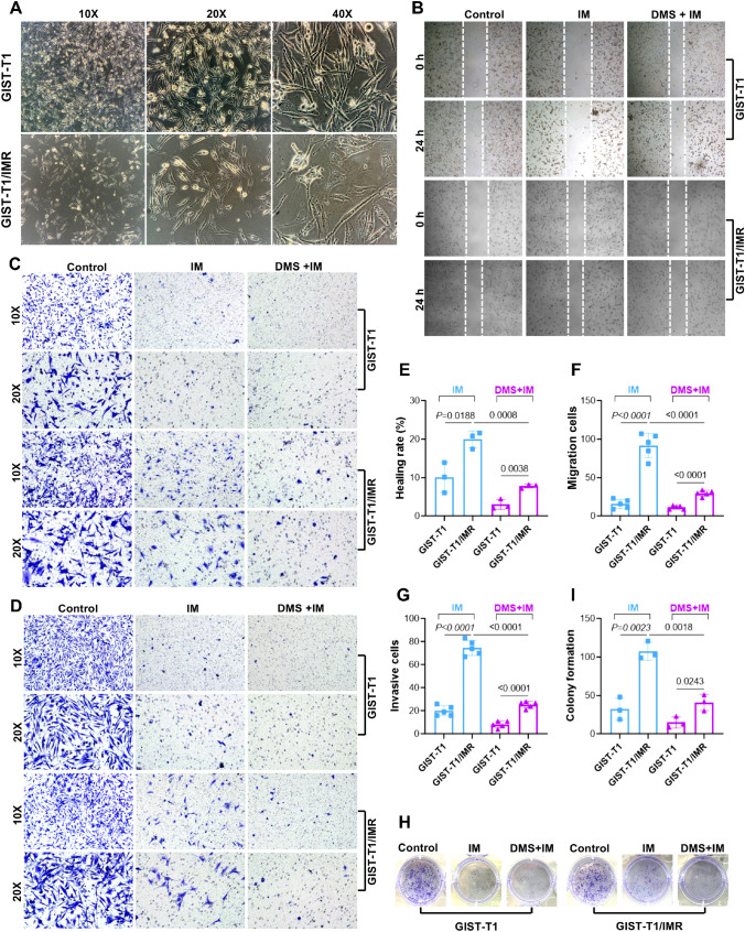
Fig. 6.
Establishment of the IM-resistant GIST-T1/IMR cell line. A Cell morphology under microscope between GIST-T1 and GIST-T1/IMR cell line. B, E. Picture of lateral cell migration and scratch healing rate between GIST-T1 and GIST-T1IR cell line. C, F Picture of longitudinal migration and migration rate between GIST-T1 and GIST-T1/IMR cell line. D, G Picture of cell transwell invasion and number of transmembrane cells. H and I. Picture of colony formation and formation rate

