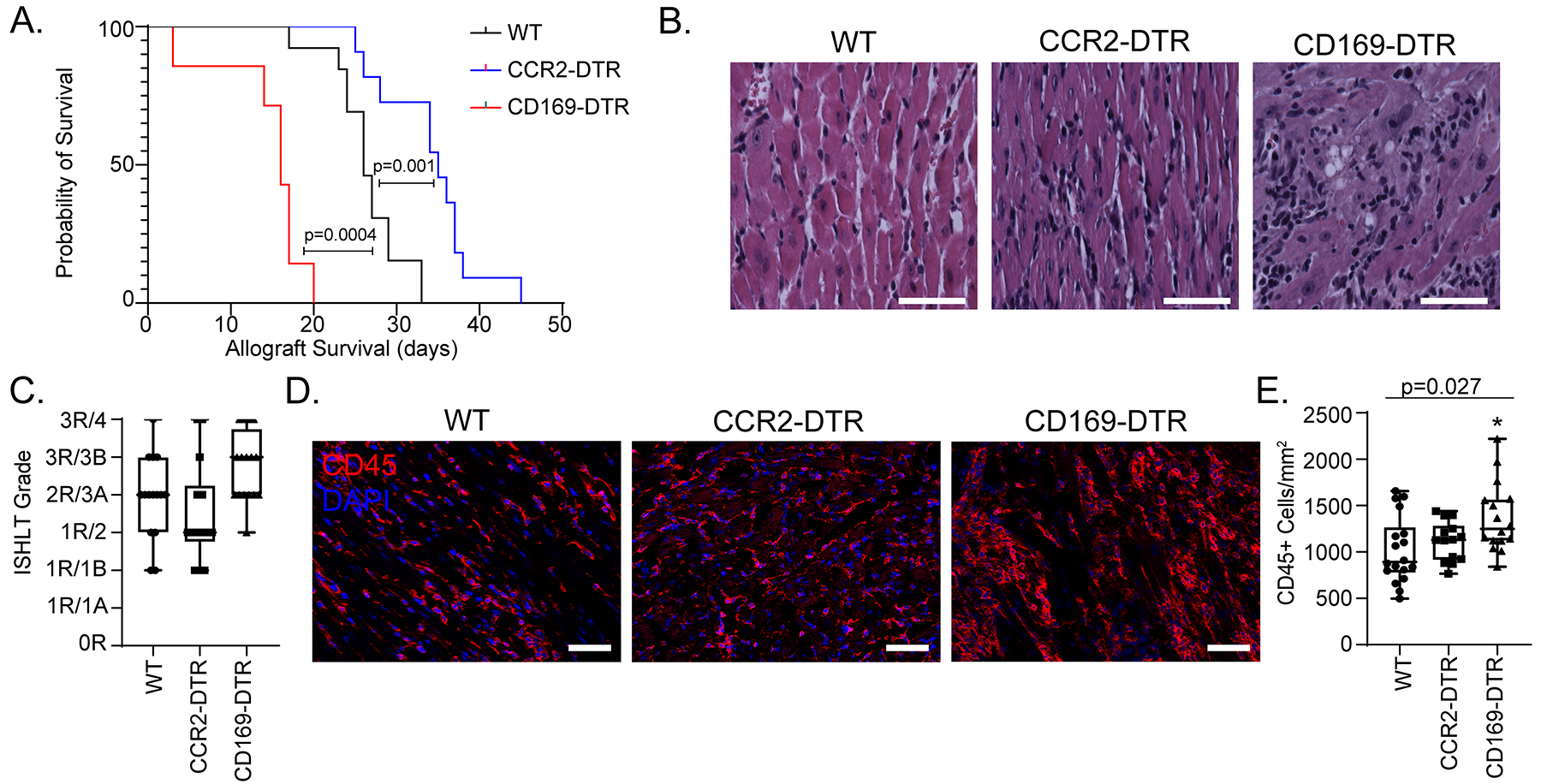
Figure 5: Donor macrophages differentially mediate allograft survival.

A) Kaplan-Meier survival curve of CD169DTR/+ (n = 7) vs CCR2DTR/+ (n = 11) vs littermate controls (n = 13) (Log-rank). B) H & E stain on transplanted hearts collected at post-transplant day 10. C) At least 3 random regions were evaluated by a trained cardiac pathologist and scored based on the 1990/2004 ISHLT cellular rejection grading guidelines (n=16 hearts for each cohort). D) Post-transplant day 10 hearts were stained with CD45 (red) and DAPI (blue). E) Number of CD45+ cell/mm2 heart was quantified. There was a significant difference among the three groups (Kruskal-Wallis; p = 0.027) and between WT and CD169DTR/+ (Dunn’s test for multiple comparisons; p=0.014). There was no difference noted between WT and CCR2DTR/+ (Dunn’s test for multiple comparisons; p = 0.58). n=16 for each cohort. Scale bar = 50 μm.
