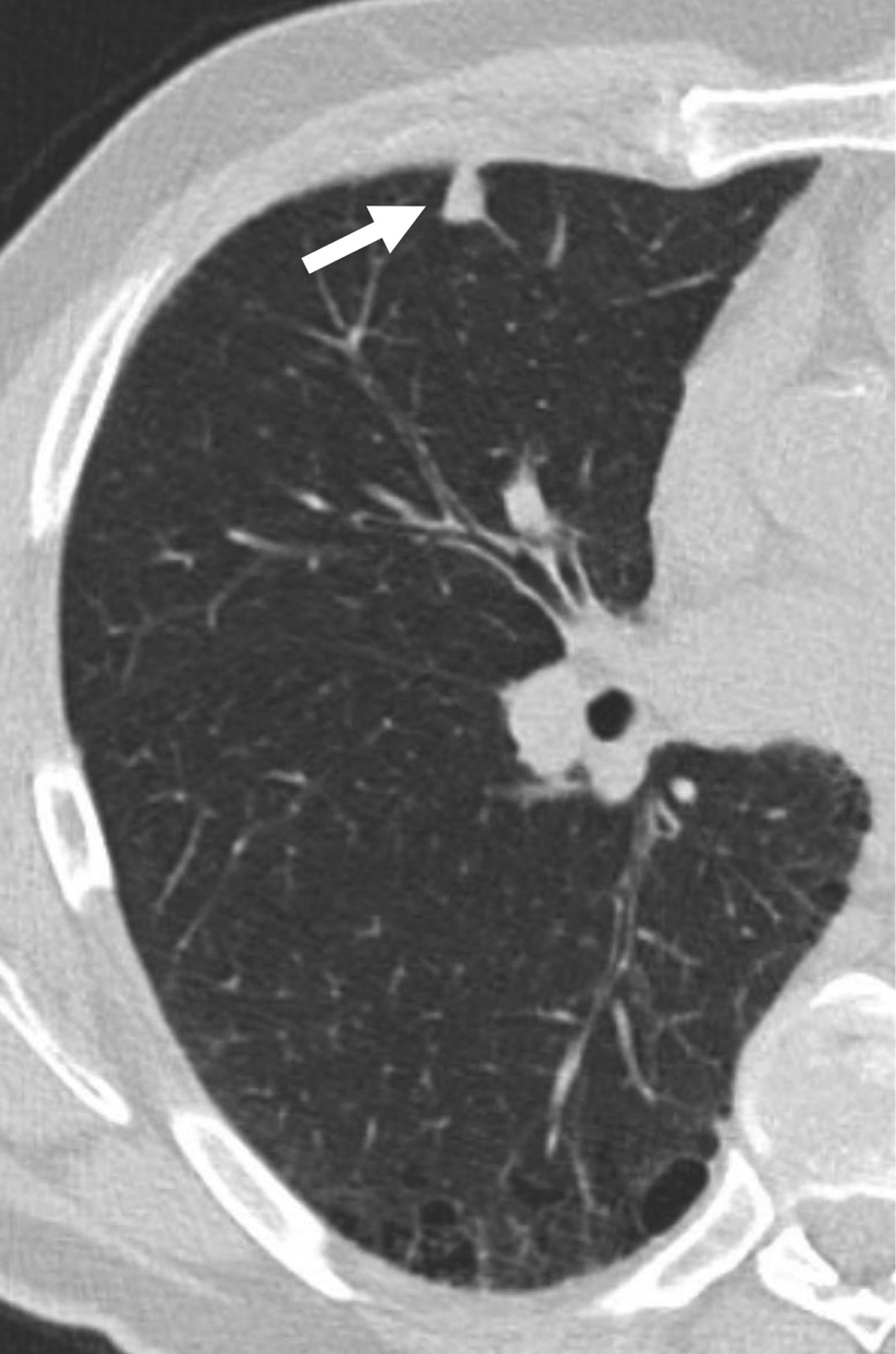
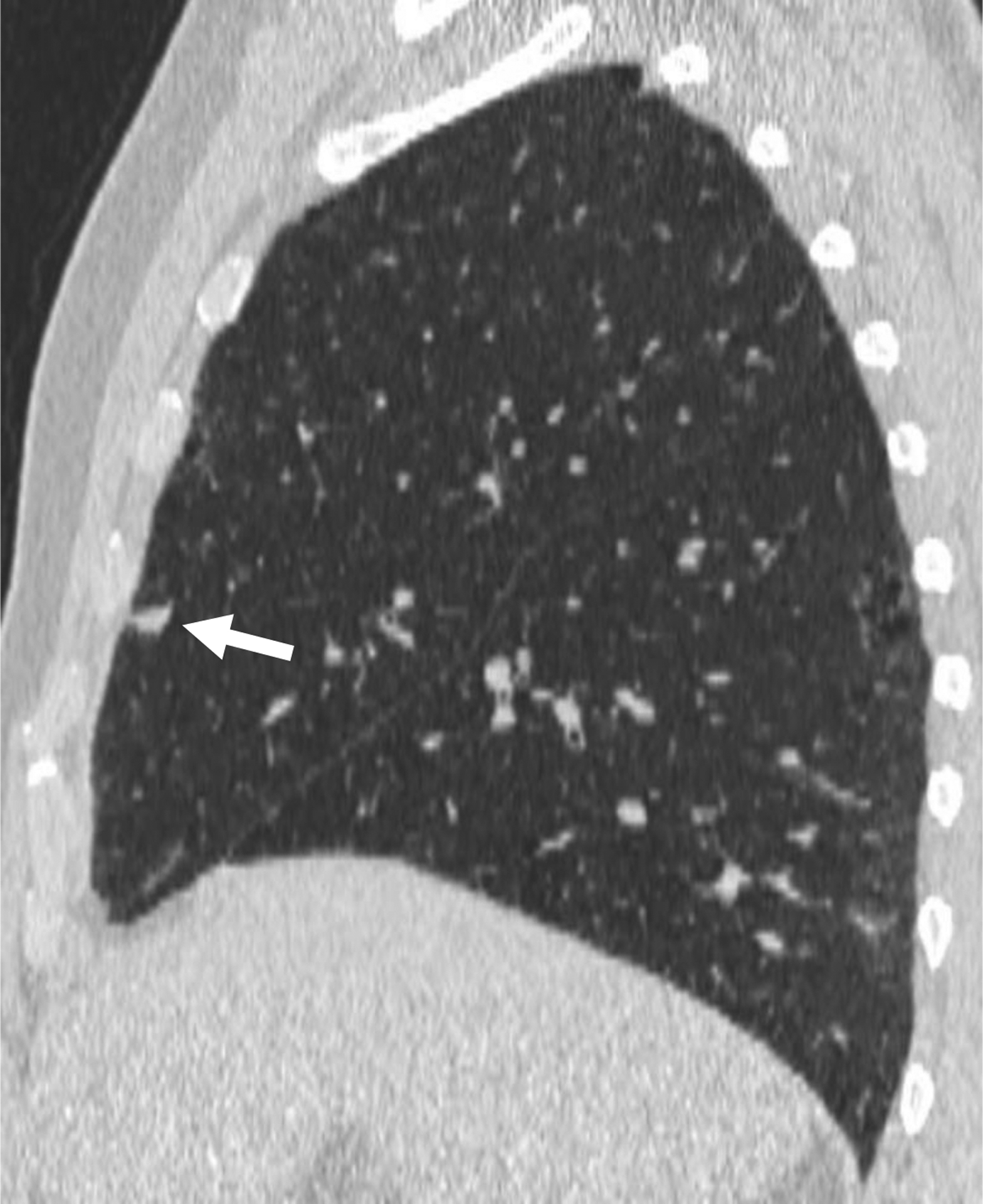
Figure 4.


66-year-old man undergoing lung cancer screening CT. (A) Axial and (B) sagittal images from baseline CT examination show a solid nodule (arrow) in the right middle lobe adjacent to an incomplete minor fissure, with mean diameter of 9.5 mm (for both readers). Both readers considered the nodule to have triangular, polygonal, or ovoid shape. Nodule was classified as category 2 by both readers given its size, perifissural location, and shape, consistent with an intrapulmonary lymph node. No cancer developed during follow-up.
