
Figure 6.
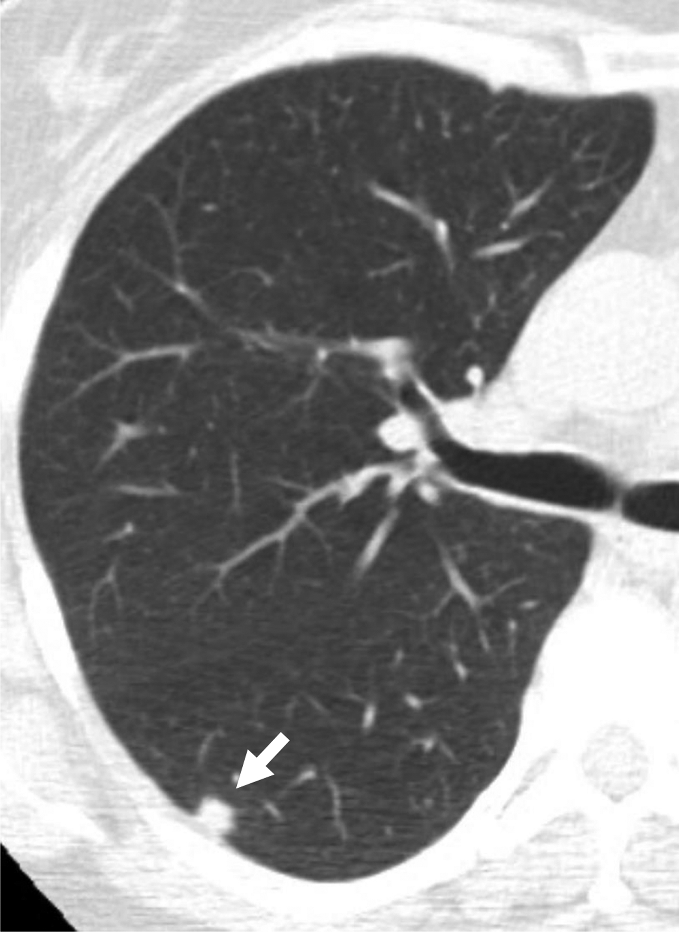

58-year-old woman undergoing lung cancer screening CT. (a) Axial image from baseline CT image shows a solid nodule (arrow) in the right lower lobe with a subpleural location (other than perifissural). Nodule had mean diameter of 8.0 mm (for both readers). Reader 2, but not reader 1, considered the nodule to have triangular, polygonal, or ovoid shape. Both readers categorized the nodule as category 3 using Lung-RADS v1.1. Using the explored modification of Lung-RADS v1.1 that considers solid nodules less than 10 mm with other subpleural location and triangular, polygonal, or ovoid shape to likely represent intrapulmonary lymph nodes, nodule remained classified as category 3 by reader 1 but was classified as category 2 by reader 2. (B) Axial image from 1-year follow-up CT examination demonstrates slight increase in size of nodule (arrow). Nodule was eventually diagnosed as lung cancer. Category 2 assignment using modified Lung-RADS by reader 2 represented a false-negative interpretation. v1.1 = version 1.1.