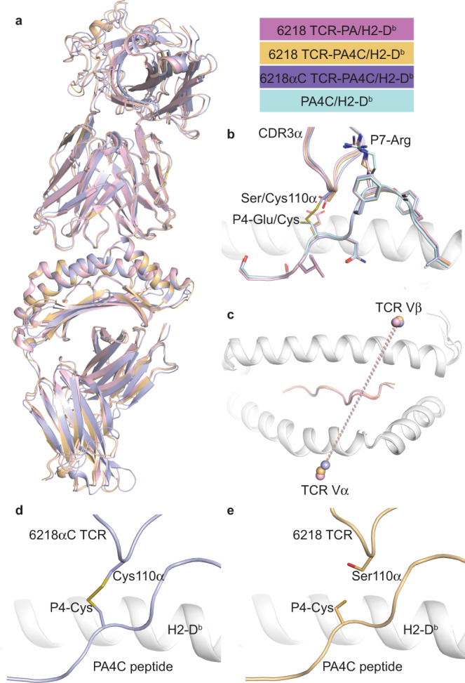
Fig. 5. TCR-peptide S-S bonding without structural rearrangement.

a Superposition of the three TCR-pMHC structures solved in this study with the 6218 TCR-PA/H2-Db complex in pink, the 6218 TCR-PA4C/H2-Db in gold, and the 6218αC TCR-PA4C/H2-Db in purple. b Zoom-in view on the TCR-peptide interface in a with the addition of the PA4C/H2-Db structure (PA4C peptide in pale blue). c Top view of the H2-Db antigen-binding cleft in white cartoon with the mass centre (sphere) of each TCR variable domain from the three TCR-pMHC structures (color coding as in a). d Structure of the 6218αC TCR (purple) in complex with PA4C/H2-Db (peptide in purple, MHC in white) showing a S-S bond formed at the interface. e Structure of the 6218 TCR (gold) in complex with PA4C peptide (gold) presented by H2-Db (white). See Supplementary Fig. 7 for electron density maps and further comparison of PA/H2-Db and PA4C/H2-Db structures.
